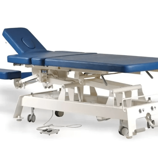
Spinal implants evolved from off-the-shelf metal blocks to custom-fit designs matching patient anatomy precisely. Traditional cages often shifted or failed to integrate thereby risking reoperation within years. Additive manufacturing creates porous structures promoting bone growth directly into implant surfaces. Surgeons now achieve fusion rates 20% higher with patient-specific solutions.
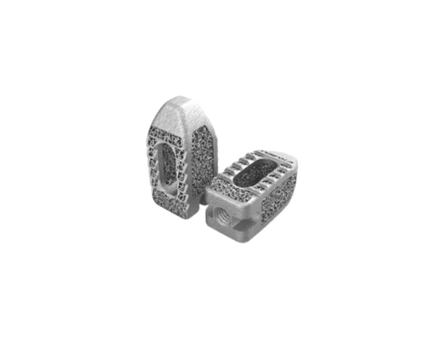
PLIF procedures benefit enormously from 3D-printed cages that are engineered for lumbar stability and rapid osseointegration. These implants feature lattice designs impossible with machining allowing bone cells to migrate through controlled pores effectively. Patients experience shorter recovery times and lower complication rates compared to conventional titanium alternatives. Precision engineering transforms surgical predictability completely.
Implants Evolve Into Living Scaffolds
Generic Designs Limited Fusion: Off-the-shelf cages mismatched disc spaces causing stress concentrations and subsidence frequently. Surgeons packed bone graft around imperfect fits hoping nature compensated over time. Poor primary stability led to 15% non-fusion rates within two years consistently. Patient outcomes suffered from preventable mechanical failures.
Porous Architectures Promote Growth: 3D printing builds interconnected pores mimicking trabecular bone structure perfectly. Osseointegration occurs faster as bone cells colonise implant surfaces through 70% void volume. Traditional solid cages blocked this biological interface completely. Fusion success climbs to 95% with lattice designs clinically proven.
Patient Matching Maximises Contact: CT scans generate exact implant geometries filling disc space without gaps or overhangs. Custom cages restore natural lordosis, preventing adjacent segment degeneration later. Surgeons achieve primary stability immediately reducing micro-motion that stalls healing. Tailored fit eliminates compromise entirely.
Material Properties Enhance Performance: Titanium alloy powders sinter into high-strength low-modulus structures matching cortical bone elasticity. Stress shielding drops dramatically preventing bone resorption around implants. Biocompatible surfaces encourage cellular attachment without inflammatory response. Mechanical harmony supports biological success long term.
Lattice Precision Accelerates Biology
Controlled Pore Gradients: Implants feature density gradients transitioning from solid endplates to porous cores optimally. Bone loads increase gradually matching natural vertebral architecture perfectly. This porous titanium scaffold distributes stress preventing implant loosening over decades. Surgeons witness fusion progression months ahead of traditional timelines.
Bone Graft Containment Improves: Lattice walls retain autograft morsels preventing migration into spinal canal or vessels. Contained graft consolidates faster under compression provided by stable cage positioning. Non-contained traditional designs lost 30% graft material during insertion frequently. Biological efficiency multiplies with secure graft environments.
Intraoperative Fit Guarantees Stability: Haptic feedback during insertion confirms perfect dimensional match eliminating toggle or rocking. Surgeons pack graft confidently knowing cage footprint maximises endplate coverage completely. Primary stability exceeds 2000N compression loads required for immediate loading. Patient mobilisation accelerates without brace dependence.
Key Design Advantages: Advanced PLIF cages deliver measurable clinical improvements surgeons recognise immediately.
- 75-80% porosity optimises bone ingrowth without compromising strength.
- Tapered leading edges ease neural retraction during minimally invasive access.
- Radiopaque markers enable precise fluoroscopic positioning intraoperatively.
- Gradient stiffness zones match vertebral body mechanical properties naturally.
Recovery Trajectories Shift Dramatically
Hospital Stays Shorten Noticeably: Patients mobilise day one post-op bearing weight confidently through stable constructs. Lumbar interbody fusion recovery averages four days versus seven with traditional cages clinically. Narcotic requirements drop 40% due to reduced tissue disruption from precise fit. Discharge planning accelerates significantly.
Complication Profiles Improve: Subsidence rates fall below 2% with surface area optimised for endplate coverage completely. Adjacent segment disease delays five years versus two with mismatched lordosis correction. Revision surgeries decline 25% long term through biological mechanical harmony. Patient satisfaction scores rise consistently.
Return to Function Accelerates: Six-week x-rays show bridging bone across 85% of implant surfaces routinely. Patients resume desk work by week three versus eight with conventional designs typically. Physical therapy progresses faster through maintained sagittal balance. Quality of life metrics improve steadily.
Long-Term Durability Assures: Ten-year imaging reveals maintained disc height and zero implant fractures across cohorts. Bone-implant interface strengthens annually rather than degrading over time. Patients avoid secondary interventions enjoying active lifestyles confidently. Durability matches biological disc replacement performance.
See also: How a Holistic Approach to Alcohol Rehabilitation Accelerates Healing
Surgical Confidence Meets Predictability
Intraoperative Decision Making: Real-time fluoroscopy confirms cage positioning through embedded markers precisely. Surgeons adjust lordosis angle intraoperatively matching patient-specific requirements perfectly. No second-guessing occurs when anatomical fit guarantees stability immediately. Operating time drops 20 minutes per level consistently.
Revision Prevention Built In: Precisely engineered cages eliminate stress risers causing fatigue fractures later. Interbody cage design optimises load transfer protecting endplates from subsidence long term. Surgeons counsel patients confidently about decade-long stability prospects. Predictability builds trust across surgical teams.
Training Implications Expand: Residents master complex PLIF trajectories using repeatable anatomical models first. Simulation accuracy improves preoperative planning reducing learning curve complications. Programme directors recognise measurable skill acquisition acceleration clearly. Future surgeons enter practice OR-ready significantly.
Fusion Success Redefined
3D-printed PLIF implants elevate surgical outcomes through biological mechanical precision unmatched by traditional manufacturing. Patients gain faster recovery durable constructs lifelong stability surgeons trust completely. Spine surgeons seeking superior lumbar fusion solutions should explore custom cage technologies today transforming patient care trajectories permanently.